IGF-1 LR3: Structure, Receptor Binding and Laboratory Use
IGF-1 LR3 started life as a production fix. In the early 1990s, an Australian team expressing IGF-1 in E. coli added a short leader sequence to help the protein fold, swapped one amino acid near the front, and ended up with an analogue that IGF-binding proteins barely recognize. Cell-culture labs have used it ever since.
This guide covers its identity and molecular weight, what each sequence change does, how it engages the IGF-1 receptor in vitro, the assays it appears in, how it compares with native IGF-1 and DES(1-3) IGF-1, and what to look for on a lot’s certificate.
For Research Use Only. Not for use in diagnostic or therapeutic procedures.
Quick answer: What is IGF-1 LR3?
IGF-1 LR3 (Long R3 IGF-I) is an 83-residue recombinant analogue of insulin-like growth factor 1, with arginine replacing glutamate at position 3 and a 13-residue N-terminal extension. These changes sharply cut its binding to IGF-binding proteins while keeping IGF-1 receptor binding. Labs use it as a serum-free media supplement and in IGF-1 receptor assays.
✓ 83-residue recombinant polypeptide, about 9.1 kDa
✓ Low IGFBP binding, with IGF-1 receptor binding kept
✓ A cell-culture and assay reagent, sold for research use only
Contents
- IGF-1 LR3 at a glance
- What is IGF-1 LR3?
- How the Arg3 and N-terminal changes affect IGFBP binding
- How IGF-1 LR3 interacts with the IGF-1 receptor
- Lab uses
- IGF-1 LR3 vs IGF-1 vs DES(1-3) IGF-1
- FDA status
- Vial storage
- Checking a batch
- FAQs
IGF-1 LR3 at a Glance
Quick answer: IGF-1 LR3 is the 70-residue IGF-1 chain with arginine at position 3 and 13 extra residues at the front, 83 residues in all and about 9.1 kDa. It binds the IGF-1 receptor, a receptor tyrosine kinase, while IGF-binding proteins hold it only weakly. Labs use it in serum-free media and receptor assays.
| Field | Value |
| Other names | Long R3 IGF-I, LR3-IGF-1 |
| Parent molecule | Insulin-like growth factor 1 (70 residues) |
| Length | 83 amino acids |
| Modifications | Glu3→Arg substitution; 13-residue N-terminal extension |
| Approx. molecular weight | ~9.1 kDa (9,111 Da calculated from the sequence with its three disulfide bonds) |
| Production | Recombinant expression |
| Receptor target | IGF-1 receptor (IGF-1R), a receptor tyrosine kinase |
| IGFBP binding | Much lower than native IGF-1 |
| Typical lab uses | Serum-free media supplement; IGF-1R binding and phosphorylation assays |
| Status | No FDA drug approval; research reagent |
| Cellugenix supply | IGF-1 LR3 1 mg, lyophilized, batch COA Purity by HPLC by HPLC |
The calculated weight comes straight from the 83-residue sequence. Different tables quote 9,111 or 9,117 Da depending on whether the three disulfide bonds are counted as closed, and the molecular formula for the reduced form is C400H625N111O115S9. For lot details, open the IGF-1 LR3 1 mg specification and COA.
What Is IGF-1 LR3?
Quick answer: IGF-1 LR3 is an engineered analogue of insulin-like growth factor 1, first described in 1992 by Francis and colleagues in Adelaide. It adds 13 residues to the N-terminus of IGF-1 and swaps glutamate 3 for arginine, giving an 83-residue polypeptide that became a standard reagent for cell-culture media.
The 13-residue leader has an unexpected origin. It’s the first 11 amino acids of methionyl porcine growth hormone followed by a Val-Asn pair, and it began as part of an E. coli fusion-protein system. The team found that this hydrophobic extension helped the protein fold correctly during production, so they kept it.
The Adelaide group’s 1992 paper in the Journal of Molecular Endocrinology described the new analogues as “very useful reagents in the investigation of IGF-I action.” The full sequence reads MFPAMPLSSLFVN, then GPR, then the remaining 67 residues of IGF-1.
What Does “LR3” Stand For?
“L” stands for Long, after the 13-residue N-terminal extension. “R3” names the arginine (single-letter code R) that replaces glutamate at position 3 of the IGF-1 chain. The 1992 paper called the molecule Long [Arg3]-IGF-I, which suppliers shortened to Long R3 IGF-I, LR3 IGF-1, and then LR3.
Is IGF-1 LR3 a Peptide?
Yes, in the broad sense: it’s an 83-residue polypeptide, long enough to count as a small protein. It folds into a defined shape held by three disulfide bonds. Unlike short peptides built by solid-phase synthesis, it’s made by recombinant expression, and Cellugenix describes its material as a recombinant IGF-1 analog.
How Do the Arg3 and N-Terminal Changes Affect IGFBP Binding?
Quick answer: In serum and conditioned media, IGF-binding proteins (IGFBPs) capture most native IGF-1. The Glu3→Arg substitution and the N-terminal extension both reduce IGFBP affinity, so far more IGF-1 LR3 stays unbound in culture. IGF-1 receptor binding is largely kept, which is why the analogue performs consistently in media.
Glutamate 3 belongs to the N-terminal stretch of IGF-1, a region that contacts the binding proteins. A companion 1992 study by King and colleagues showed that swapping it for arginine left an analogue that “bound very poorly” to IGFBP-2, while its receptor binding dropped only slightly. The extension adds bulk near the same surface and lowers IGFBP contact further.
Cell lines secrete their own IGFBPs into the medium, and serum adds more. Native IGF-1 gets soaked up by those proteins, so the free fraction that can reach a receptor changes with cell density, medium age and serum lot. IGF-1 LR3 largely escapes that capture, keeping the free concentration close to what was added.
The 1992 data make the point neatly. In a fibroblast line that secretes no detectable IGFBPs, Long R3 IGF-I was less potent than native IGF-1, so its edge appears only when binding proteins are present.
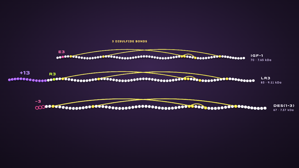
Comparing IGF-1 and IGF-1 LR3 sequences, showing the Arg3 substitution and N-terminal extension
How Does IGF-1 LR3 Interact With the IGF-1 Receptor?
Quick answer: IGF-1 LR3 binds the IGF-1 receptor, a receptor tyrosine kinase. Binding triggers receptor autophosphorylation and recruitment of IRS-1, which activates the PI3K-Akt and MAPK/ERK signaling pathways. These phosphorylation events are the readouts most IGF-1R assays measure in cultured cells.
The IGF-1 receptor is a pre-formed dimer: two α subunits outside the cell that bind the ligand, linked to two β subunits that span the membrane and carry the tyrosine kinase domain. In a 2018 review, Fumihiko Hakuno and Shin-Ichiro Takahashi of the University of Tokyo summarized the first step: “Ligand binding to the α subunit of the receptor leads to a conformational change.”
From there, the cascade runs in a fixed order:
- Binding: IGF-1 LR3 docks on the α subunits.
- Autophosphorylation: the β-subunit kinases phosphorylate each other’s tyrosine residues.
- Docking: IRS-1 and SHC bind the phosphorylated receptor and are phosphorylated in turn.
- Branching: IRS-1 recruits the p85 subunit of PI3K, starting the PI3K-Akt branch, while GRB2 links to Ras and the MAPK/ERK branch.
Each step leaves a measurable mark. Phospho-IGF-1R, phospho-IRS-1, phospho-Akt and phospho-ERK can all be detected by ELISA or western blot, which is how labs confirm that a ligand lot is active.
What Is IGF-1 LR3 Used For in the Lab?
Quick answer: IGF-1 LR3 is used mainly as a supplement in serum-free cell-culture media, including CHO and other production cell lines, and as a ligand in IGF-1 receptor assays. Commercial grades range from research grade and “receptor grade” to GMP grade for manufacturing media, with specifications for purity, endotoxin and activity.
| Assay or use | What it measures | Typical readout |
| Serum-free media supplementation | Cell-line growth in defined media | Cell count, viability |
| Receptor binding assay | Ligand binding to IGF-1R | Competitive binding, Kd |
| Receptor phosphorylation assay | IGF-1R activation | Phospho-IGF-1R by ELISA or western blot |
| Downstream signaling assay | Pathway activation | Phospho-Akt, phospho-ERK |
| Cell proliferation assay | Ligand activity in vitro (ED50) | BrdU incorporation, MTT |
In cell culture and binding assays, IGF-1 LR3 also works as a tool for separating receptor effects from IGFBP effects. Running it beside native IGF-1 in the same cells shows how much of a response depends on binding-protein capture.
Why Biomanufacturing Uses It
Serum-free production media need a defined growth factor that stays available. A 2000 study in Biotechnology Progress compared insulin with LongR3 in two CHO cell lines making recombinant proteins and found LongR3 better sustained viability under production conditions. The authors concluded “LongR(3) is the preferred growth factor for cell lines VA12 and CL23.”
IGF-1 LR3 vs IGF-1 vs DES(1-3) IGF-1
Quick answer: Native IGF-1, IGF-1 LR3 and DES(1-3) IGF-1 are all IGF-1 receptor ligands. They differ in length and in how strongly IGF-binding proteins capture them, which changes how much stays free in culture. IGF-1 LR3 adds residues to the front, and DES(1-3) IGF-1 removes the first three.
| IGF-1 | IGF-1 LR3 | DES(1-3) IGF-1 | |
| Length | 70 aa | 83 aa | 67 aa |
| Change | None (reference) | Glu3→Arg + 13-aa extension | First 3 residues (Gly-Pro-Glu) removed |
| IGFBP binding | High | Much reduced | Reduced |
| Typical lab use | Reference ligand | Media supplement, IGF-1R assays | Signaling and IGFBP studies |
DES(1-3) IGF-1 is a natural variant, originally purified from bovine colostrum. A 1996 profile by the same Adelaide group traced its low IGFBP binding to “the absence of the glutamate at position 3,” the same residue the LR3 design replaces. The two analogues reach a similar result by different routes: one deletes glutamate 3, the other swaps it for arginine.
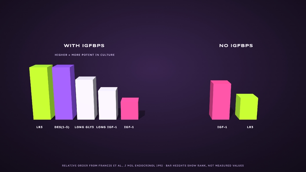
Ranking of IGF-1 analogue potency in cell lines with and without IGF-binding proteins
Is IGF-1 LR3 FDA-Approved?
Quick answer: IGF-1 LR3 has never been approved by the FDA as a drug. Labs receive it as an in-vitro research reagent, and its label reads “For Research Use Only. Not for use in diagnostic or therapeutic procedures.” That status applies to every lot Cellugenix supplies.
Research reagents in the US are defined by their labeling and intended use, a point covered in our guide to research peptides and US law. For the label wording itself, see what Research Use Only means.
Storage of the Sealed Vial
Each vial holds freeze-dried (lyophilized) IGF-1 LR3 under an intact seal, with the lot number printed on its label. Keep it sealed, out of the light and away from humidity, following the conditions that label gives. Temperature ranges are listed in the storage and handling guide.
How to Verify Research-Grade IGF-1 LR3
Quick answer: The batch certificate of analysis comes first. The IGF-1 LR3 1 mg lot on sale was tested by Testides, an independent lab, on January 5, 2026, and reached Purity by HPLC by HPLC-UV, with measured content alongside. The report number links back to the lab’s own database, so anyone can confirm the document.
| Field on the certificate | Current IGF-1 LR3 1 mg value |
| Report number | CP-IGF-1-122925 (opens the same record on testides.com) |
| Purity and method | 97.7% by HPLC-UV |
| Content | Measured milligrams in the vial, checked against the 1 mg label |
| Tested | January 5, 2026 |
| Laboratory | Testides, which operates separately from Cellugenix |
The certificate covers purity and content. Labs that need identity by mass or activity data for a specific assay can run those checks in-house by LC-MS, MALDI or SDS-PAGE. The live record is linked from the IGF-1 LR3 1 mg certificate page, and the walkthrough titled how to read a COA explains every field. The two analytical methods are compared in HPLC vs mass spectrometry.
Frequently Asked Questions
What is IGF-1 LR3?
IGF-1 LR3 is an engineered analogue of insulin-like growth factor 1 with 83 residues: the 70-residue IGF-1 chain, arginine in place of glutamate at position 3, and a 13-residue N-terminal extension. Those changes lower binding to IGF-binding proteins, so labs use it in serum-free media and IGF-1 receptor assays.
What does LR3 stand for?
LR3 is short for Long R3. “Long” refers to the 13-residue extension added to the N-terminus, which began as part of an E. coli fusion system. “R3” refers to arginine, single-letter code R, at position 3 of the IGF-1 chain, where native IGF-1 carries glutamate. The original 1992 name was Long [Arg3]-IGF-I.
What is the molecular weight of IGF-1 LR3?
IGF-1 LR3 weighs about 9.1 kDa. Calculated from its 83-residue sequence with three disulfide bonds closed, the figure is 9,111 Da, and tables that count the reduced form list about 9,117 Da. The lot’s certificate of analysis reports purity and content; mass measurement would need a separate method such as LC-MS.
How is IGF-1 LR3 different from native IGF-1?
IGF-1 LR3 has 13 more residues and an arginine at position 3. Native IGF-1 binds IGF-binding proteins tightly, so much of it is captured in serum or conditioned media. IGF-1 LR3 escapes most of that binding while still engaging the IGF-1 receptor, which keeps its free concentration steadier in culture.
Is IGF-1 LR3 a peptide or a protein?
Both labels fit. At 83 residues it falls right on the boundary, and many sources call it a small protein or polypeptide. It folds into a defined three-dimensional structure held by three disulfide bonds, and it’s made by recombinant expression, the route used for proteins, where short peptides are usually built by chemical synthesis.
Is IGF-1 LR3 FDA-approved?
No. There’s no FDA approval of IGF-1 LR3 for any purpose. Cellugenix stocks it strictly as an in-vitro lab reagent, with a label reading “For Research Use Only. Not for use in diagnostic or therapeutic procedures.” Its documented uses are serum-free cell-culture media and IGF-1 receptor assays.
The Bottom Line
IGF-1 LR3 is an 83-residue IGF-1 analogue built for low IGF-binding-protein capture, which makes it a dependable serum-free media supplement and IGF-1 receptor assay ligand. Its value depends on the presence of binding proteins in the system. Purity by HPLC appears on every batch certificate.
IGF-1 LR3 1 mg specification and COA
For Research Use Only. Not for use in diagnostic or therapeutic procedures.
Sources
- Francis GL et al. Journal of Molecular Endocrinology, 1992; 8(3): 213–223. PMID 1378742.
- King R et al. Journal of Molecular Endocrinology, 1992; 8(1): 29–41. PMID 1311930.
- Hakuno F, Takahashi SI. Journal of Molecular Endocrinology, 2018; 61(1): T69–T86. PMID 29535161.
- Morris AE, Schmid J. Biotechnology Progress, 2000; 16(5): 693–697. PMID 11027158.
- Ballard FJ et al. International Journal of Biochemistry & Cell Biology, 1996; 28(10): 1085–1087. PMID 8930132.
For Research Use Only. Not for use in diagnostic or therapeutic procedures.